Physiology - Comparison between mammals and birds

Comparison between mammals and birds


Table 1. Comparison of number of cones, visual information process, and proportion of nerve fibres that do not cross over to the opposite side in the optic chiasm between mammal and avian


Mammal
Bird
Number of cones
Most diurnal mammals have 2 cones (dichromatic).
Diurnal birds have 3 to 4 types. Most birds are tetrachromatic.

The cones in birds contain small, coloured oil droplets which absorb certain wavelengths before light reaches the photopigment.

This “light filtration” results the sensitivity of the cones in more finely tuned to the optimal wavelength than in mammals.
Visual information process
Occurs in the paired nuclei superior colliculus in the midbrain.

In mammals, the optic tectum is a small sized part in the midbrain.
Occurs in the optic tectum in the midbrain.

In birds, the optic tectum is a dominant part of the midbrain.
Proportion of nerve fibres that do not cross over to the opposite side in the optic chiasm
Human: 50%
Cat: 30%
Dog: 25%
Horse: 15%

Almost 100%

Reference
  1. Sjaastad O.V., Sand O. and Hove K. (2010) Physiology of domestic animals, 2nd edn., Oslo: Scandinavian Veterinary Press.

Physiology - Visual pathway

Visual pathway

1. Visual sensory pathway is initiated primarily in the retina and ends in the primary visual cortex of the brain.

Figure 1: The rod pathway. A large number of rods are converging on a single rod bipolar cell (RBC) with A17 and AII amacrine cells modulating the output. DCB and HCB are depolarizing and hyperpolarizing cone bipolar cells, respectively, that output to ganglion cells (GC). Their input and output are modulated by horizontal cells (HC) and AII amacrine cells, respectively. 
Source: Maggs D.J., Miller P.E. and Ofri R. (2013) Slatter's fundamentals of veterinary ophthalmology, 5th edn., Missouri: Elsevier.


Figure 2. Plain of the retinal layers. All 10 cellular and synaptic layers are indicated.
Source: Maggs D.J., Miller P.E. and Ofri R. (2013) Slatter's fundamentals of veterinary ophthalmology, 5th edn., Missouri: Elsevier.

2. Initiation of the visual pathway and process of the visual features are initiated by the photoreceptor cells (retinal neurons) responding to light and contrast.
  • Photoreceptor cells are connected to ganglion cells via series of bipolar cells. The distal part of rods and cones (located in the photoreceptor layer and outer nuclear layer) are connected with the proximal part of bipolar cells and horizontal cells (located in the outer plexiform layer) and ganglion cells (located in the inner plexiform layer and ganglion layer) are connected with the distal part of bipolar cells and amacrine cells (located in the inner plexiform layer).
Figure 3. Antagonism of central and peripheral receptive fields of a bipolar or ganglion cell. (a) In an on-centre-off-surround cell, the cell is activated by a light stimulating its centre and inhibited when the light is turned off. (b) The same cell is inhibited by a light stimulating its surround and activated when the surround light is turned off. (c) When both centre and surround are illuminated, the net result is moderate activation as the centre is excited and the surround is inhibited. Same principle applies to light off situation. (d)
Opposite responses are observed in an off-centre-on-surround cell. The cell is inhibited by a centre stimulating light and activated when the light is turned off. (e) Activation of the same cell occurs when the surround light is turned on and inhibited when is is turned off. (f) When both centre and surround are illuminated (or in dark), the net result is moderate activation as the centre is inhibited and the surround is excited.
Source: Maggs D.J., Miller P.E. and Ofri R. (2013) Slatter's fundamentals of veterinary ophthalmology, 5th edn., Missouri: Elsevier.

3. The signal activity is passed on to the optic nerve formed by the axons of the ganglion cells in the retina.
  • Ganglion cells have receptive field responsible for responding to light stimulus. Each receptive field is composed of two regions, a centre and a surround, each responding oppositely to the light source. 
  • In case of “on-centre cell response”,if the light exposure is on the centre of receptive field, centre illumination will be increased and surround illumination will be decreased due to stimulation of on-centre cell and inhibition of the same cell via light exposure on the surround of receptive field.
  • In case of “off-centre response”, if the light exposure is on the surround of receptive field, centre illumination will be decreased and surround illumination will be increased due to stimulation of off-centre cell and inhibition of the same cell via light exposure on the centre of receptive field.
    • The most common neurotransmitter released as a response to the light stimulation is 'Glutamate' which works as the bipolar cell inhibitory. 
    • On-centre cell response and off centre cell response are determined by the neural circuits in the retina:
    1. Straight through pathways


      • The rods and cones are synaptically connected to two types of bipolar cells (on and off cells).
      • Steps: On centre bipolar cells and on centre off surround ganglion cells are used as an example.
        1. Presence of light in the centre of receptive field.
        2. Hyperpolarisation of the centre cones.
        3. Less glutamate (inhibitory neurotransmitter for on centre bipolar cell) is released at the synapse.
        4. Hence on-centre bipolar cell is depolarised as less bipolar inhibitory was released.
        5. Increased amount of glutamate is released at the synapse due to depolarisation.
        6. This will cause depolarisation of the on centre ganglion cell.
      • Off centre bipolar cells will result in hyperpolarisation of the on centre ganglion cells due to different receptors at the synapse of the bipolar cells and causing an opposite response. 
    2. Lateral pathways
       

      • The connection of centre cones and surround cones cones in the fovea with via acmacrine and horizontal cells. 
      • Antagonistic effect on the centre of receptive field
      • Occurs as the eye is trying to focus on the cencentrated (strong stimulation received in the surround) information rather than the information received in the centre which is not as concentrated as the surround information.
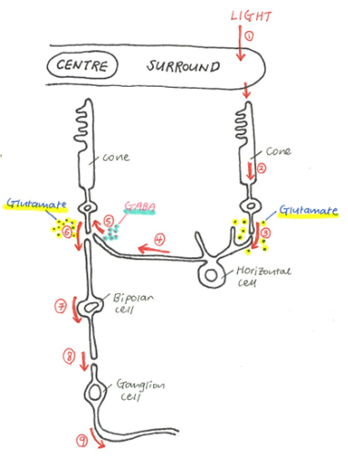
      • Steps: On centre bipolar cells and off centre on surround ganglion is used as an example
        1. Presence of light in the surround receptive field and darkness in the centre receptive field.
        2. Hyperpolarisation of the surround cones and depolarisation of the centre cones. 
        3. Hyperpolarisation of the surround will release less neurotransmitter (glutamate) at the synapse.
        4. Hence the horizontal cells are hyperpolarised. 
        5. Stimulus on the horizontal cells will cause release of neurotransmitter called Gamma Aminobutyric Acid (GABA) at the synapse with centre cone and this will have no impact on the production of glutamate by the centre cone as hyperpolarisation of horizontal cells will not produce much of GABA.
        6. Large amount of glutamate is released at the synapse (due to depolarisation of the centre cones) 
        7. On centre bipolar cells are hyperpolarised as glutamate acts as an inhibitory neurotransmitter. 
        8. Decreased amount of glutamate is released at the synapse due to hyperpolarisation.
        9. This will cause hyperpolarisation of the on centre ganglion cells.
    • Function of neurotransmitters:
        • Glutamate
            • Excitatory neurotransmitter for horizontal cell
            • Inhibitory neurotransmitter for on centre bipolar cell
            • Excitatory neurotransmitter for off centre bipolar cell
            • Excitatory neurotransmitter for both on and off centre ganglion cell
        • GABA (Gamma Aminobutyric Acid)
            • Inhibitory neurotransmitter for neighbouring cones


Figure 4. The primary visual pathway. The primary visual pathway entailing the retina, optic nerves, optic chiasm, optic tract, hypothalamus, dorsal lateral geniculate nucleus, optic radiation, and primary visual cortex. Source:Dale P. (2010) Brains how they work and what they tells us about who we are, 1st edn., New Jersey: Pearson Education.

4. The signal passes through the partial crossing of axons at the optic chiasm (partial decussation) where left and right visual information delivered via the optic nerve cross to the opposite side of the lateral geniculate nucleus via the optic tract.
  • The medial half (nasal half) of each retina receives light rays from the lateral portion of the visual field and crosses to the opposite lateral geniculate nucleus.
  • The lateral half (temporal half) of each retina receives light rays from the medial portion of the visual field and remains to the same lateral geniculate nucleus.
  • The right optic tract carries signals representing the left half of the visual field.
  • The left optic tract carries signals from the right visual field.

5. All the axons synapse at the lateral geniculate nucleus and spread through the brain as the geniculostriate radiation.

6. The signal travels to the primary visual cortex in the occipital lobe.
  • Receptive fields of neuron in the primary visual cortex include two kinds of neurons:
    • Simple cells
      • Specific rotation axis required for visual stimulus. Non specific axis will have no stimulus.
      • Receptive fields include: dark bar, light bar and light-dark bar.
    • Complex cells
      • Specific rotation axis of bars and edges required for visual stimulus. Non specific axis will have stimulus but less.
      • Non specific to the positions of stimulatory bars and edges.
References
  1. Akers R.M. and Denbow D.M. (2013) Anatomy and physiology of domestic animals, 2nd edn., Iowa: John Wiley & Sons, Inc.
  2. Aughey E. and Frye E.L. (2001) Comparative veterinary histology, 1st edn., London: Manson Publishing Ltd.
  3. Dale P. (2010) Brains how they work and what they tells us about who we are, 1st edn., New Jersey: Pearson Education.
  4. James G.C. and Bradley G.K. (2007) Cunningham's textbook of veterinary physiology, 5th edn., St. Louis: Saunders Elsevier.
  5. Maggs D.J., Miller P.E. and Ofri R. (2013) Slatter's fundamentals of veterinary ophthalmology, 5th edn., Missouri: Elsevier.
  6. Sjaastad O.V., Sand O. and Hove K. (2010) Physiology of domestic animals, 2nd edn., Oslo: Scandinavian Veterinary Press.

Physiology - Eye movement

Eye movement

Rapid eye movement
  • Eyes move with rapid jerks (saccadic movements) when looking around
  • Generates a continuous series of images of different areas
  • Eyes fixed on individual areas for 0.2-0.6 seconds before next rapid eye movement
  • Surrounding is perceived to be stable when the brain interprets this continuous series of images
  • Points of fixation are not evenly distributed but are concentrated around contours and distinctive features

Slow eye movement
  • Allow focus on moving object when head is stationary or focus on stationary objects when animal is moving
  • Vestibulo-ocular reflex
    • Fix image on retina when head is moving
    • Ocular muscles move eyeballs simultaneously but in opposite direction  to head movements
  • Optokinetic reflex
    • Stabilise moving image on retina when head is stationary
    • Rapid saccade brings eye back to middle position just before slow eye movement reaches outer limit

References
  1. Sjaastad O.V., Sand O. and Hove K. (2010) Physiology of domestic animals, 2nd edn., Oslo: Scandinavian Veterinary Press.

Physiology - Adaptation to light and dark

Adaptation to light and dark

Light adaptation
  • Adaptation to light > adaptation to dark
    • Recovers normal vision in less than a minute
  • Pupil constriction à restrict amount of light entering eye
  • Bleaching of photopigments
  • In bright light:
    • Sensory cells contain little uncleaved photopigment
    • Low light sensitivity
    • sensory cells are light adapted

Dark adaptation


Figure 1: Dark adaptation curves. Purple: rod adaptation curve. Green: cone adaptation curve. Red: two-stage dark adaptation curve of both rods and cones. The initial time delay between "light-adapted sensibility" and beginning of the curves is due to delay between time the lights are switched off and when measurement of curves start.
Source: Goldstein E.B. (2014) Sensation and Perception, 9th edn., Wadsworth: Cengage Learning.

  • Photopic (light-adapted) state to scotopic (dark-adapted) state
  • Maximal light sensitivity
    • Cones adapt more quickly to darkness than rods
      • Cones: 5 minutes
      • Rods: 20-30 minutes
    • Species dependent
    • Dependent on pre-existing light level
      • Brighter pre-existing light, lower rhodopsin stores à longer to reach maximal light sensitivity
  • Increase light sensitivity by
    • Dilation of pupil
    • Synaptic adaptation of retinal neurons
    • Increase rhodopsin available in rod outer segments à regeneration of rod photopigments

References
  1. Akers R.M. and Denbow D.M. (2013) Anatomy and physiology of domestic animals, 2nd edn., Iowa: John Wiley & Sons, Inc.
  2. Goldstein E.B. (2014) Sensation and Perception, 9th edn., Wadsworth: Cengage Learning.
  3. Maggs D.J., Miller P.E. and Ofri R. (2013) Slatter's fundamentals of veterinary ophthalmology, 5th edn., Missouri: Elsevier.
  4. Sjaastad O.V., Sand O. and Hove K. (2010) Physiology of domestic animals, 2nd edn., Oslo: Scandinavian Veterinary Press.

Physiology - Colour vision

Colour vision


Figure 1: Visual spectrum at different wavelengths. Humans can only perceive 390nm to 700nm on the visual spectrum. 
Source: Genoa College of Fine Arts (2014) Chromatology, Available at: http://www.accademialigustica.it/blog/?page_id=58 (Accessed: 3rd June 2014).
  • Colour of object is determined by which wavelengths of light is absorbed or reflected.
    • White objects = reflect all wavelengths
    • Black objects = absorb all wavelengths
    • Red object = absorbs more of the shortwave part of the spectrum & long wave light is reflected
  • Different type of cones with maximal sensitivity to light of different wavelengths = basis for colour vision
  • Birds & lower vertebrates: at least 3 different types of cones = EXCELLENT colour vision
  • Mammals (including all domestic animals): 2 types of cones
    • Exception: some primates & humans
  • Few species: 1 type
  • Primates (including human) : developed 3rd type
    • 3 types of cones in primates absorb light most efficiently in blue, green-yellow & orange-red parts of the spectrum
    • Blue, geen and red cones
    • Overlap between wavelengths absorbed by these 3 cones
    • Human brain can discriminate between colours
      • ie. 560nm: green & red stimulated equally & blue not affected = yellow
  • Colour vision based on 
    • 3 types of cones = Trichromatic
    • 2 types of cones = Dichromatic

Figure 2: Three different types of cones found in trichromatic primates, including humans, have different sensitivities to light of different wavelengths. Thus forming the basis for colour vision in these primates.
Source: Sjaastad O.V., Sand O. and Hove K. (2010) Physiology of domestic animals, 2nd edn., Oslo: Scandinavian Veterinary Press.

Fun facts

  • In vertebrates, it has been found that animals must discriminate between stimuli before expressing preference for a colour
    • e.g. Frogs forced to choose between two adjacent, illuminated panels. They select the bluer one
  • Fish have good colour vision
    • Fish retinas contain multiple classes of photopigments for colour vision
    • e.g Gold fish absorbency spectrum shifted to longer wavelengths
  • Primate colour vision spectral range: 400-700nm. 
  • Diurnal birds have a larger range than primates!
  • Red-green colour blindness
    • Caused by absence of long or middle wavelength-sensitive visual photopigments (specifically cones)
    • Been proven that addition of a third cone class can produce improved trichromatic vision in primates


References
  1. Genoa College of Fine Arts (2014) Chromatology, Available at: http://www.accademialigustica.it/blog/?page_id=58 (Accessed: 3rd June 2014).
  2. Jacobs G. H. (1983) 'Colour vision in animals', Endeavour, 7(3), pp. 137-140.
  3. Mancuso K., Hausworth W.W., Li Q., Connor T.B., Kuchenbecker J.A., Mauck M.C., Neitz J. and Neitz M. (2009) ' Gene therapy for red-green colour blindness in adult primates', Nature, 461, pp. 784-787.
  4. Sjaastad O.V., Sand O. and Hove K. (2010) Physiology of domestic animals, 2nd edn., Oslo: Scandinavian Veterinary Press.
Physiology - Rods and cones

Rods and cones
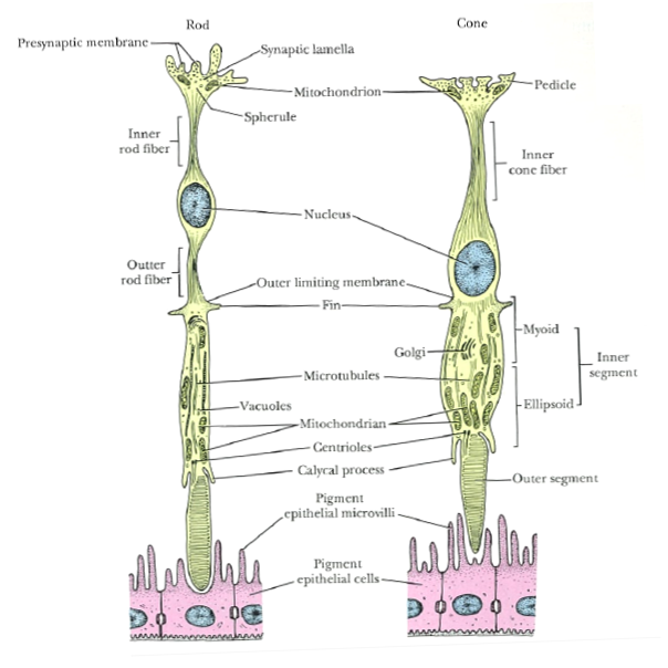
Rods and cones are the two major types of sensory cells in the eye and are located in the outer most later of the retina, closest to the choroid.


Figure 1: Diagram of rod and cone cells. Outer segments of rods and cones are closely associated with adjacent pigment epithelium.
Source: Ross M.H. and Pawlina W. (2006) Histology a text and atlas with correlated cell and molecular biology, 5th edn., Baltimore: Lippincott Williams & Wilkins.

Rods
  • Do not provide colour vision
  • Extremely sensitive
  • In poor light conditions à all vertebrae see in black, white and grey
  • In very bright conditions à lose ability to discriminate between light intensities
Cones
  • Colour vision
  • Stimulated only in good light conditions i.e. level at which rods are maximally stimulated
  • Ability to provide detailed vision in full daylight (photopic vision)
  • Area centralis of diurnal species have high number of cones
Humans

  • Density of cones are high in the fovea in the middle of the area centralis
  • Fovea in humans contain only cones à visual acuity in fovea is high
  • As fovea lack rods, area is not stimulated in weak light
  • Rod density is highest in area immediately adjacent to area centralis
Animals
  • Many species, including cattle and horses, lack a circular area centralis
  • Have visual streak
    • Density of sensory cells is high
    • Elongated region that corresponds to the projection of the horizon on retina

Table 1: Summary of the characteristics of rods and cones

Characteristics of rods and cones
Rods
Cones
Function in low light levels (scotopic)
Function in high light levels (photopic)
Sensitive to small change in light intensity
Insensitive to small change in light intensity
Low visual discrimination (low acuity)
High visual discrimination (high acuity)
Responsive to blue light
Responsive to red light
No colour differentiation
·         Contain only 1 photopigment
Colour differentiation
·         In species with 2 or more cone populations (defined by photopigments)
Sensitive to motion
Sensitive to contrast
Detect light flashing at low frequency
Detect light flashing at high frequency
More in peripheral area
More in central retina


References
  1. Maggs D.J., Miller P.E. and Ofri R. (2013) Slatter's fundamentals of veterinary ophthalmology, 5th edn., Missouri: Elsevier.
  2. Ross M.H. and Pawlina W. (2006) Histology a text and atlas with correlated cell and molecular biology, 5th edn., Baltimore: Lippincott Williams & Wilkins.
  3. Sjaastad O.V., Sand O. and Hove K. (2010) Physiology of domestic animals, 2nd edn., Oslo: Scandinavian Veterinary Press.

Physiology - Light stimulation of sensory cells

Light stimulation of sensory cells
  • When light strikes the retina, photons are trapped by the receptor molecules (rods and cones) located in the membrane discs in the outer segments
  • Membrane discs in rod = "light trap"
  • Receptor molecules in retina = photopigments
  • Rods contain photopigment, rhodopsin
  • Different types of cones have its own type of photopigments
  • Photopigments compose of opsin (protein) and retinal (produced in cell from vitamin A, retinol)
  • Photopigments are G-protein-coupled receptors


Figure 1: Cleavage of photopigments and transduction in rods and cones due to light stimulation. This causes a chemical bond change, resulting in a straightened molecule. Retinal detaches from opsin, causing conformational change in its shape. G-protein transducin binds to opsin and is activated. Hyperpolarization of the cell and reduced release of neurotransmitter ends the chain reaction. 
Source: Sjaastad O.V., Sand O. and Hove K. (2010) Physiology of domestic animals, 2nd edn., Oslo: Scandinavian Veterinary Press.

Darkness
  • Outer segment of rods and cones:
    • Concentration of cGMP in outer segment is high
    • Surface membrane:
      • High density of Na+ channels so Na+ can diffuse into cells.
      • Channels open when bound to cGMP
      • Ion channels open in dark à Na+ enters & depolarize sensory cells (light)
  • Inner segment of rods and cones:
    • Na+ pumped out via Na+-K+ pump 
    • Depolarization of inner segment keeps voltage-gated Ca2+ channels open à continuous neurotransmitter (glutamate) release from synaptic terminal of sensory cell
Light
  • When light (photons) is absorbed, 11-cis-retinal of the photopigment, rhodopsin, changes its conformational shape to all-trans-retinal
  • All-trans-retinal leaves photopigment, causing a conformational change of the photopigment
  • Now, there is a exposed binding receptor site on photopigment
  • G-protein transducin can now attach to the photopigment and is activated
  • This protein structure activates the enzyme, phosphodiesterase
  • Phosphodiesterase hydrolyses cGMP resulting in
  • closure of Na+ channels 
  • Na+ concentration in cell decreases and membrane potential = -ve
  • Rods & cones hyperpolarize
  • Release of neurotransmitter, Glutamate, is reduced
  • Results in inhibition & stimulation of ganglion cells
Figure 2: Ion currents through rods and cones in darkness. Na+ diffuses into the outer segment through open ion channels while Na+ is pumped out via Na+K+ pump in the inner segment. Light closes the ion channels.
Source: Sjaastad O.V., Sand O. and Hove K. (2010) Physiology of domestic animals, 2nd edn., Oslo: Scandinavian Veterinary Press.

Notes
  • Single photon can close many hundreds of ion channels
  • Prevents more than a million Na+ from entering cell
  • Single photon evokes detectable receptor potential in cell
Reference
  1. Sjaastad O.V., Sand O. and Hove K. (2010) Physiology of domestic animals, 2nd edn., Oslo: Scandinavian Veterinary Press.

Physiology - Image formation

Image formation
Formation of a clear image is achieved through three important processes: light refraction, accommodation and pupil diameter.

Light refraction

Figure 1: Image formation in the eye. Light refraction at the air-cornea interface is greater than refraction at the aqueous humour-lens inferface.
Source: Sjaastad OV, Sand O, Hove K. 2010. Physiology of domestic animals. 2nd ed. Oslo: Scandinavian Veterinary Press.

  • Cornea and lens refracts light
  • Refraction at air-cornea interface > refraction at aqueous humour-lens interface
    • Cornea responsible for most of eye's refractive power
  • Inverted image (upside down) focused onto rods and cones in retina

Accommodation

Lens changes shape and increase refractive power to focus objects at different distances on the retina.
Figure 2: Accomodation. a: Ciliary body is relaxed when viewing distant objects. Zonular fibres are taut and lens is flattened. b: Ciliary body is contracted when viewing nearby objects. Zonular fibres are relaxed and lens is more spherical.
Source: Sjaastad O.V., Sand O. and Hove K. (2010) Physiology of domestic animals, 2nd edn., Oslo: Scandinavian Veterinary Press.
Far vision
  • Ciliary muscles relaxed
  • Zonular fibres tightened
  • Lens flattened
  • Focus distant object on retina

Near vision
  • Ciliary muscles contracted
  • Zonular fibres relaxed
  • Lens becomes more spherical and convex (natural resting shape of lens)
  • Refraction of light increases à focus nearby object on retina

Pupil diameter

Image 3: Variation in diameter of pupil depending on light intensity. Radial muscle fibres contract in dim light while circular muscle fibres contract in strong light.
Source: Sjaastad O.V., Sand O. and Hove K. (2010) Physiology of domestic animals, 2nd edn., Oslo: Scandinavian Veterinary Press.

  • Controls amount of light entering eye
  • Controlled by circular muscle fibers of sphincter pupillae muscle and radial muscle fibres of radial dilator pupillar muscle
Dim light
  • Increased activity in sympathetic nerve fibres à radial muscle contraction
  • Pupil dilates à allow more light to enter eye 

Strong light
  • Increased stimulation by parasympathetic nerve fibres à circular muscle contraction
  • Pupil narrows à  restrict amount of light entering eye
  • Constriction prevents divergent light rays from entering eye à rays fall on periphery of retina where they would not be focused properly

References
  1. Akers R.M. and Denbow D.M. (2013) Anatomy and physiology of domestic animals, 2nd edn., Iowa: John Wiley & Sons, Inc.
  2. Dyce K.M., Sack W.O. and Wensing C.J.G. (2010) Textbook of veterinary anatomy, 4th edn., Missouri: Saunders Elsevier.
  3. Sjaastad O.V., Sand O. and Hove K. (2010) Physiology of domestic animals, 2nd edn., Oslo: Scandinavian Veterinary Press.