
Common diseases
Conjunctivitis


Ocular squamous cell carcinoma

Figure 1: Limbal squamous cell carcinoma in a horse. Carcinoma is seen as a light pink lobulated mass on the limbus extending to the cornea and conjunctiva.
Source: Hendrix D.V.H. (2005) 'Equine Ocular Squamous Cell Carcinoma', Clinical Techniques in Equine Practice, 4(1), pp. 87-94.
 Figure 2: Third eyelid replaced by squamous cell carcinoma in a horse.
Source: Hendrix D.V.H. (2005) 'Equine Ocular Squamous Cell Carcinoma', Clinical Techniques in Equine Practice, 4(1), pp. 87-94.
 Figure 3: Ulcerated squamous cell carcinoma in the medial canthus of a horse.
Source: Hendrix D.V.H. (2005) 'Equine Ocular Squamous Cell Carcinoma', Clinical Techniques in Equine Practice, 4(1), pp. 87-94.


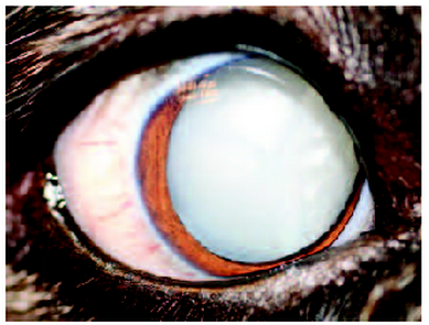
Figure 5: Mature senile cataract of a dog.
Source: Mitchell N. (2006) 'Treatment of canine cataracts using phacoemulsification Part 1: When to refer', Companion Animal, 11(3), pp. 83-87.
Conjunctivitis
Figure 1: Allergic conjunctivitis with hyperemia and mucoid discharge in a dog.
Source: Gelatt K.N. (2008) Essentials of veterinary ophthalmology, 2nd edn., USA: Blackwell Publishing.
Figure 2: Viral conjunctivitis in a dog with canine distemper.
Source: Gelatt K.N. (2008) Essentials of veterinary ophthalmology, 2nd edn., USA: Blackwell Publishing.
Figure 3: Viral conjunctivitis with chemosis and hyperemia in a cat with feline herpesvirus.
Source: Gelatt K.N. (2008) Essentials of veterinary ophthalmology, 2nd edn., USA: Blackwell Publishing.
- Inflammation of conjunctival membrane
- Red eye with discharge
- Serous discharge
- Physical irritants
- Allergy
- Viral agents
- Treatment: Flush eye with sterile saline eyewash or artificial tears 3-4 times a day
- Mucoid discharge
- Follicles on underside of third eyelid
- Eye irritant
- Infection
- Treatment: Antibiotic and corticosteroid eye ointments
- Purulent discharge
- Infection
- May crust eyelids
- Treatment: Remove mucus and pus from eyes and apply antibiotics
- Bilateral - allergy or systemic disease
- Unilateral - foreign body in the eye
- Clinical signs
- Hyperemia
- Chemosis
- Ocular discharge
- Harmorrhage
- Follicle formation
- Pruritis
- Pain/discomfort
- Cats: twitching
- Dogs: no discomfort
- Classification
- Infectious
- Bacterial
- Dogs: usually caused by Streptococcus and Staphylococcus
- Cats: Chlamydophila Felis, Bordetella bronchiseptica
- Viral
- Dogs: canine distemper virus
- Cats: feline herpesvirus-1, Feline calicivirus
- Fungal
- Rickettsial
- Rickettsia rickettsii
- Parasitic
- Cats: Thelazia californiensis
- Noninfectious
- Allergic
- Follicular
- Cats
- Lipogranulomatous
- Eosinophilic
Ocular squamous cell carcinoma
Source: Hendrix D.V.H. (2005) 'Equine Ocular Squamous Cell Carcinoma', Clinical Techniques in Equine Practice, 4(1), pp. 87-94.
Source: Hendrix D.V.H. (2005) 'Equine Ocular Squamous Cell Carcinoma', Clinical Techniques in Equine Practice, 4(1), pp. 87-94.
Source: Hendrix D.V.H. (2005) 'Equine Ocular Squamous Cell Carcinoma', Clinical Techniques in Equine Practice, 4(1), pp. 87-94.
Figure 4: Limbal squamous cell carcinoma in a cattle.
Source: Tsujita H. and Plummer C.E. (2010) 'Bovine Ocular Squamous Cell Carcinoma',Veterinary Clinics of North America: Food Animal Practice, 26(3), pp. 511-526.
Figure 5: Corneal squamous cell carcinoma in a cattle.
Source: Tsujita H. and Plummer C.E. (2010) 'Bovine Ocular Squamous Cell Carcinoma',Veterinary Clinics of North America: Food Animal Practice, 26(3), pp. 511-526.
- Tumour of the eye and ocular adnexa
- Nictitating membrane/third eyelid
- Conjunctiva
- Eyelids
- Cornea
- Limbus
- Cattle
- 7-9 years old
- Herefold
- Kept in subtropcal areas - environmentally favourable
- White faced and pink skin around eyes
- Incidence: 0.8-5%
- Holstein
- Whole-carcass slaughter
- 80% of bovine tumors reported at slaughter
- Pigs, goats and sheep - low incidence
- Horses
- 10-11 years old
- Lack of periocular pigmentation and ultraviolet light exposure
- Draft breeds
- Colorado
- Belgians
- Clydesdales
- Light breeds
- Appaloosa
- Albino
- Colour dilute
- Clinical signs
- Conjunctiva or limbus: small white to light pink mass
- Limbal to corneal: multilobulated and compacted
- Eyelid: ulcerative mass
- Treatment
- Surgical excision
- Cryosurgery
- Radiofrequency Hyperthermia
- Irradiation therapy
- Radiotherapy
- Laser ablation
- Immunotherapy
- Topical and intralesional chemotherapy
Uveitis
Figure 1. Uveitis of a cow
Source: Bruce H.G., Cheryl L.C. and Robert L.P. (2004) Veterinary ophthalmology essentials, 1st edn., Philadelphia: Elsevier.
Figure 2. Uveitis of a guinea pig
Source: Bruce H.G., Cheryl L.C. and Robert L.P. (2004) Veterinary ophthalmology essentials, 1st edn., Philadelphia: Elsevier.
Figure 3. Uveitis of a horse
Source: Bruce H.G., Cheryl L.C. and Robert L.P. (2004) Veterinary ophthalmology essentials, 1st edn., Philadelphia: Elsevier.
Figure 4. Uveitis of a rabbit
Source: Bruce H.G., Cheryl L.C. and Robert L.P. (2004) Veterinary ophthalmology essentials, 1st edn., Philadelphia: Elsevier.
- Inflammation of the uveal tract: iris, ciliary body and choroid
- Anterior uveitis: Iris and ciliary body
- Posterior uveitis: choroid and retina
- Inflammation mediated by vascular impairment
- Animals with uveitis are susceptible to secondary infection
- Causes
- Infection
- Parasites
- Fungi
- Bacteria
- Trauma
- Metabolic disease
- Cancer
- Tumor
- Clinical signs
- Blepharospasm
- Conjunctival hypermia
- Chemosis
- Corneal edema
- Aqueous flare (proteins and cells leaking into anterior chamber due to impaired blood-aqueous barrier)
- Miosis (small sized pupil)
- Unique clinical signs in cats
- Nodular accumulation of inflammatory cells within the iris storma
- Treatment
- Must be treated aggressively to prevent further development (e.g. glaucoma, scarring or loss of vision)
- Aspirin
- Corticosteroid- injection under the eyelid, eyedropper or oral medication
- Atropine
Cataracts
Figure 4: Cataracts in a foal.
Source: Brooks D.E. (2002) 'Equine Ophtalmology', American Association of Equine Practitioners Proceedings, 48, pp. 300-313.
Figure 5: Mature senile cataract of a dog.
Source: Mitchell N. (2006) 'Treatment of canine cataracts using phacoemulsification Part 1: When to refer', Companion Animal, 11(3), pp. 83-87.
- Opacity in the lens of the animals giving the animal a blurry vision
- Vacuoles in equatorial region
- Proceeding to lens nucleus becoming more opaque and spreading to the entire lens
- Occurrence due to
- Disease
- Old age
- Trauma to the eye
- Inheritance
- Can be produced by diets deficient in riboflavin or tryptophane
- Clinical signs
- Change in colour of the eye
- Behaviour of animal changed due to increasing loss of vision i.e clumsiness
- Treatment
- Surgery
- Phacoemulsification cataract surgery = high success rate in foals
- Topical antibiotics after surgery:
- Chloramphenicol, gentamicin, ciproflaxacin, tobramycin ophthalmic solutions
- 1% atropine = maintain blood-aqueous barrier, minimize pain from ciliary muscle spasm and dilation of pupils.
- Corticosteroid = prevent inflammation
References
- Brooks D.E. (2002) 'Equine Ophtalmology', American Association of Equine Practitioners Proceedings, 48, pp. 300-313.
- Bruce H.G., Cheryl L.C. and Robert L.P. (2004) Veterinary ophthalmology essentials, 1st edn., Philadelphia: Elsevier.
- Eldredge D.M., Carlson L.D., Carlson D.G. and Giffin J.M. (2007) Dog owner's home veterinary handbook, 4th edn., New Jersey: Howell Book House.
- Gelatt K.N. (2008) Essentials of veterinary ophthalmology, 2nd edn., USA: Blackwell Publishing.
- Hendrix D.V.H. (2005) 'Equine Ocular Squamous Cell Carcinoma', Clinical Techniques in Equine Practice, 4(1), pp. 87-94.
- King T.C., Priehs D.R., Gum G.G. and Miller T.R. (1991) 'Therapeutic management of ocular squamous cell carcinoma in the horse: 43 cases (1 979-1 989)', Equine Veterinary Journal, 23(6), pp. 449-452.
- Kinoshita J.H., Kador P. and Catiles M. (1981) 'Aldose reductase in diabetic cataracts', The Journal of American Medical Association, 246(3), pp. 257-261.
- Mitchell N. (2006) 'Treatment of canine cataracts using phacoemulsification Part 1: When to refer', Companion Animal, 11(3), pp. 83-87.
- Richter C.P. and Duke J.R. (1970) 'Cataracts produced in rats by yoghurt', American Association for the Advancement of Science, 168(3937), pp. 1372-1374.
- Tsujita H. and Plummer C.E. (2010) 'Bovine Ocular Squamous Cell Carcinoma',Veterinary Clinics of North America: Food Animal Practice, 26(3), pp. 511-526.
- Turner S.M. (2008) Small animal ophthalmology, 1st edn., Saunders Elsevier.