Showing posts with label clinical signs. Show all posts

Pathology - Common diseases

Common diseases

Conjunctivitis

Figure 1: Allergic conjunctivitis with hyperemia and mucoid discharge in a dog.
Source: Gelatt K.N. (2008) Essentials of veterinary ophthalmology, 2nd edn., USA: Blackwell Publishing.

Figure 2: Viral conjunctivitis in a dog with canine distemper.
Source: Gelatt K.N. (2008) Essentials of veterinary ophthalmology, 2nd edn., USA: Blackwell Publishing.

Figure 3: Viral conjunctivitis with chemosis and hyperemia in a cat with feline herpesvirus.
Source: Gelatt K.N. (2008) Essentials of veterinary ophthalmology, 2nd edn., USA: Blackwell Publishing.

  • Inflammation of conjunctival membrane
    • Red eye with discharge
      • Serous discharge
        • Physical irritants
        • Allergy 
        • Viral agents
        • Treatment: Flush eye with sterile saline eyewash or artificial tears 3-4 times a day
      • Mucoid discharge
        • Follicles on underside of third eyelid
        • Eye irritant
        • Infection
        • Treatment: Antibiotic and corticosteroid eye ointments
      • Purulent discharge
        • Infection
        • May crust eyelids
        • Treatment: Remove mucus and pus from eyes and apply antibiotics
    • Bilateral - allergy or systemic disease
    • Unilateral - foreign body in the eye
  • Clinical signs
    • Hyperemia
    • Chemosis
    • Ocular discharge
    • Harmorrhage
    • Follicle formation
    • Pruritis
    • Pain/discomfort
      • Cats: twitching
      • Dogs: no discomfort
  • Classification
    • Infectious
      • Bacterial
        • Dogs: usually caused by Streptococcus and Staphylococcus
        • Cats: Chlamydophila Felis, Bordetella bronchiseptica
      • Viral
        • Dogs: canine distemper virus
        • Cats: feline herpesvirus-1, Feline calicivirus
      • Fungal
      • Rickettsial
        • Rickettsia rickettsii
      • Parasitic
        • Cats: Thelazia californiensis
    • Noninfectious
      • Allergic
      • Follicular
    • Cats
      • Lipogranulomatous
      • Eosinophilic

Ocular squamous cell carcinoma


Figure 1: Limbal squamous cell carcinoma in a horse. Carcinoma is seen as a light pink lobulated mass on the limbus extending to the cornea and conjunctiva.
Source: Hendrix D.V.H. (2005) 'Equine Ocular Squamous Cell Carcinoma', Clinical Techniques in Equine Practice, 4(1), pp. 87-94.

Figure 2: Third eyelid replaced by squamous cell carcinoma in a horse.
Source: Hendrix D.V.H. (2005) 'Equine Ocular Squamous Cell Carcinoma', Clinical Techniques in Equine Practice, 4(1), pp. 87-94.

Figure 3: Ulcerated squamous cell carcinoma in the medial canthus of a horse.
Source: Hendrix D.V.H. (2005) 'Equine Ocular Squamous Cell Carcinoma', Clinical Techniques in Equine Practice, 4(1), pp. 87-94.


Figure 4: Limbal squamous cell carcinoma in a cattle.
Source: Tsujita H. and Plummer C.E. (2010) 'Bovine Ocular Squamous Cell Carcinoma',Veterinary Clinics of North America: Food Animal Practice, 26(3), pp. 511-526.

Figure 5: Corneal squamous cell carcinoma in a cattle.
Source: Tsujita H. and Plummer C.E. (2010) 'Bovine Ocular Squamous Cell Carcinoma',Veterinary Clinics of North America: Food Animal Practice, 26(3), pp. 511-526.
  • Tumour of the eye and ocular adnexa
    • Nictitating membrane/third eyelid
    • Conjunctiva
    • Eyelids
    • Cornea
    • Limbus
  • Cattle
    • 7-9 years old
    • Herefold
      • Kept in subtropcal areas - environmentally favourable
      • White faced and pink skin around eyes
      • Incidence: 0.8-5%
    • Holstein
    • Whole-carcass slaughter
      • 80% of bovine tumors reported at slaughter
  • Pigs, goats and sheep - low incidence
  • Horses
    • 10-11 years old
    • Lack of periocular pigmentation and ultraviolet light exposure
    • Draft breeds
      • Colorado
      • Belgians
      • Clydesdales 
    • Light breeds
      • Appaloosa
      • Albino
      • Colour dilute
  • Clinical signs
    • Conjunctiva or limbus: small white to light pink mass
    • Limbal to corneal: multilobulated and compacted
    • Eyelid: ulcerative mass
  • Treatment
    • Surgical excision
    • Cryosurgery
    • Radiofrequency Hyperthermia
    • Irradiation therapy
    • Radiotherapy
    • Laser ablation
    • Immunotherapy
    • Topical and intralesional chemotherapy

Uveitis

Figure 1. Uveitis of a cow
Source: Bruce H.G., Cheryl L.C. and Robert L.P. (2004) Veterinary ophthalmology essentials, 1st edn., Philadelphia: Elsevier.

Figure 2. Uveitis of a guinea pig
Source: Bruce H.G., Cheryl L.C. and Robert L.P. (2004) Veterinary ophthalmology essentials, 1st edn., Philadelphia: Elsevier.

Figure 3. Uveitis of a horse
Source: Bruce H.G., Cheryl L.C. and Robert L.P. (2004) Veterinary ophthalmology essentials, 1st edn., Philadelphia: Elsevier.

Figure 4. Uveitis of a rabbit
Source: Bruce H.G., Cheryl L.C. and Robert L.P. (2004) Veterinary ophthalmology essentials, 1st edn., Philadelphia: Elsevier.
  • Inflammation of the uveal tract: iris, ciliary body and choroid
    • Anterior uveitis: Iris and ciliary body
    • Posterior uveitis: choroid and retina
  • Inflammation mediated by vascular impairment
  • Animals with uveitis are susceptible to secondary infection
  • Causes
    • Infection
      • Parasites
      • Fungi
      • Bacteria
    • Trauma
    • Metabolic disease
    • Cancer
    • Tumor
  • Clinical signs
    • Blepharospasm
    • Conjunctival hypermia
    • Chemosis
    • Corneal edema
    • Aqueous flare (proteins and cells leaking into anterior chamber due to impaired blood-aqueous barrier)
    • Miosis (small sized pupil)
    • Unique clinical signs in cats
      • Nodular accumulation of inflammatory cells within the iris storma
  • Treatment
    • Must be treated aggressively to prevent further development (e.g. glaucoma, scarring or loss of vision)
    • Aspirin
    • Corticosteroid- injection under the eyelid, eyedropper or oral medication
    • Atropine

Cataracts


Figure 4: Cataracts in a foal.
Source: Brooks D.E. (2002) 'Equine Ophtalmology', American Association of Equine Practitioners Proceedings, 48, pp. 300-313.


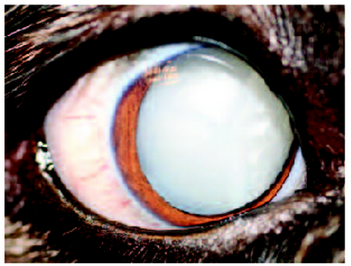
Figure 5: Mature senile cataract of a dog. 
Source: Mitchell N. (2006) 'Treatment of canine cataracts using phacoemulsification Part 1: When to refer', Companion Animal, 11(3), pp. 83-87.

  • Opacity in the lens of the animals giving the animal a blurry vision
  • Vacuoles in equatorial region
  • Proceeding to lens nucleus becoming more opaque and spreading to the entire lens
  • Occurrence due to
    • Disease
    • Old age
    • Trauma to the eye
    • Inheritance
    • Can be produced by diets deficient in riboflavin or tryptophane
  • Clinical signs
    • Change in colour of the eye
    • Behaviour of animal changed due to increasing loss of vision i.e clumsiness
  • Treatment
    • Surgery
      • Phacoemulsification cataract surgery = high success rate in foals
    • Topical antibiotics after surgery:
      • Chloramphenicol, gentamicin, ciproflaxacin, tobramycin ophthalmic solutions
      • 1% atropine = maintain blood-aqueous barrier, minimize pain from ciliary muscle spasm and dilation of pupils. 
      • Corticosteroid = prevent inflammation
References
  1. Brooks D.E. (2002) 'Equine Ophtalmology', American Association of Equine Practitioners Proceedings, 48, pp. 300-313.
  2. Bruce H.G., Cheryl L.C. and Robert L.P. (2004) Veterinary ophthalmology essentials, 1st edn., Philadelphia: Elsevier.
  3. Eldredge D.M., Carlson L.D., Carlson D.G. and Giffin J.M. (2007) Dog owner's home veterinary handbook, 4th edn., New Jersey: Howell Book House.
  4. Gelatt K.N. (2008) Essentials of veterinary ophthalmology, 2nd edn., USA: Blackwell Publishing.
  5. Hendrix D.V.H. (2005) 'Equine Ocular Squamous Cell Carcinoma', Clinical Techniques in Equine Practice, 4(1), pp. 87-94.
  6. King T.C., Priehs D.R., Gum G.G. and Miller T.R. (1991) 'Therapeutic management of ocular squamous cell carcinoma in the horse: 43 cases (1 979-1 989)', Equine Veterinary Journal, 23(6), pp. 449-452.
  7. Kinoshita J.H., Kador P. and Catiles M. (1981) 'Aldose reductase in diabetic cataracts', The Journal of American Medical Association, 246(3), pp. 257-261.
  8. Mitchell N. (2006) 'Treatment of canine cataracts using phacoemulsification Part 1: When to refer', Companion Animal, 11(3), pp. 83-87.
  9. Richter C.P. and Duke J.R. (1970) 'Cataracts produced in rats by yoghurt', American Association for the Advancement of Science, 168(3937), pp. 1372-1374.
  10. Tsujita H. and Plummer C.E. (2010) 'Bovine Ocular Squamous Cell Carcinoma',Veterinary Clinics of North America: Food Animal Practice, 26(3), pp. 511-526.
  11. Turner S.M. (2008) Small animal ophthalmology, 1st edn., Saunders Elsevier.

Pathology - Ruminants

Ruminants

Keratoconjunctivitis (Pink-eye)

Figure 1. Infectious bovine keratoconjunctivitis in a calf. Note the corneal vascularization, stromal abscess, and scars.
Source: Bruce H.G., Cheryl L.C. and Robert L.P. (2004) Veterinary ophthalmology essentials, 1st edn., Philadelphia: Elsevier.

  • Found in sheep, goats and calves
  • Primary infection occurs by Chlamydiae
  • Chlamydiae: Gram negative, obligatory intra cellular parasites
  • Cytoplasmatic inclusion bodies in corneal cells found in the infected animals
  • Clinical signs
    • Watery discharge from the eye
    • Clouding of the cornea
    • Secondary bacterial infection is common due to mechanical damage of the cornea and altered local defence system allowing potential pathogenic bacteria to contribute to secondary infections
  • Treatment
    • Vaccination
      • Penicillins, spectinomycin or other aminocyclitols do not respond
      • Tetracyclines respond
    • High and steady plasma levels of suitable antibiotics for more than one week

Thelazia rhodesi (Eye Worms)

File:Thelazis01.jpg
Figure 2: Presence of Thelazia. 
Source: University of Sydney Centre for Veterinary Education (2012) Thelazia spp, Available at:http://vetbook.org/wiki/cow/index.php/Thelazia_spp (Accessed: 3rd June 2014).

  • Parasite of domestic animals, wild animals and less commonly horses
  • Occurs at surface of cornea, near nictitating membrane, in conjunctival sac and lachrymal duct
  • Transmission occurs through route of flies that feed on animal's ocular secretions, tears and conjunctiva.
  • Clinical signs
    • Conjunctivitis
    • Keratitis
    • Lacrimation
    • Ocular discharge
    • Ulcers
    • Corneal opacity
    • Epiphora - excessive tears production
  • Treatment
    • Mechanical removal of worms using forceps after putting anesthesia 
    • Topical treatment with antiparasitic drugs applied to eyes directly
    • Irrigate with aqueous solution of Boric powder
    • Concurrent use with Ciplox-D (Ciprofloxacin and dexamethasone) eye drops every 8 hours for 5 days 
      • suppression of inflammation & secondary bacterial infections.
    • Ivermectin to be injected subcutaneously @ 0.2mg/Kg bodyweight
    • Fly control

Reference
  1. Bogaard A.E.J.M. (2011) 'Veterinary Quarterly', Inclusion keratoconjunctivitis ('pink eye') in sheep, 6(4), pp. 229 -235.
  2. Bruce H.G., Cheryl L.C. and Robert L.P. (2004) Veterinary ophthalmology essentials, 1st edn., Philadelphia: Elsevier.
  3. Deepthi B. and Yalavarthi C. (2012) 'Eye Worm infection in a Cattle - A Case Report', Veterinary World, 5(4), pp. 236-237.
  4. Naem S. (2006) 'Thelazia rhodesi (Spirurida,  Thelaziidae), bovine eyeworm: morphological study by scanning electron microscopy', Parasitol Research, 100, pp. 855-860.
  5. Otranto D., Tarsitano E., Tarversa D., Giangaspero A., DeLuca F. and Puccini V. (2001) 'Differentiation among three species of bovine Thelazia (Nematoda: Thelaziidae) by polymerase chain reaction–restriction fragment length polymorphism of the first internal transcribed spacer ITS-1 (rDNA)', International Journal of Parasitlogy, 31(14), pp. 1693-1698.
  6. Otranto D. and Traversa D. (2005) 'Thelazia eyeworm: an original endo- and ecto- parasitic nematode' Trends in Parasitology, 21(4), pp. 1-4.
  7. University of Sydney Centre for Veterinary Education (2012) Thelazia spp, Available at:http://vetbook.org/wiki/cow/index.php/Thelazia_spp (Accessed: 3rd June 2014).

Pathology - Companion animals

Companion animals

Cherry eye or Haws

Figure 1: Unilateral prolapse of third eyelid gland in an American Cocker spaniel dog.
Source: Mehdi M., Sepehr M., Omid, Kazemi H. and Elnaz (2012) 'Clinical evaluation of the pocket technique for replacement of prolapsed gland of the third eyelid in dogs',Turkish Journal of Veterinary and Animal Sciences, 36(4), pp. 352-356.

Figure 2: Bilateral prolapse of third eyelid gland in a mixed-breed dog.
Source: Mehdi M., Sepehr M., Omid, Kazemi H. and Elnaz (2012) 'Clinical evaluation of the pocket technique for replacement of prolapsed gland of the third eyelid in dogs',Turkish Journal of Veterinary and Animal Sciences, 36(4), pp. 352-356.

Figure 3: Unilateral prolapse of third eyelid in a Burmese cat.
Source: Chahory S., Crasta M., Trio S. and Clerc B. (2004) 'Three cases of prolapse of the nictitans gland in cats', Veterinary Ophthalmology, 7(6), pp. 417-419.

Figure 4: Bilateral prolapse of third eyelid in a Persian cat.
Source: Chahory S., Crasta M., Trio S. and Clerc B. (2004) 'Three cases of prolapse of the nictitans gland in cats', Veterinary Ophthalmology, 7(6), pp. 417-419.

  • Prolapse of the third eyelid gland
    • Unilateral or bilateral
    • Unclear cause
    • Secondary to inflammation
    • Weakness in connective tissue that anchors the gland
      • May be heritable
  • Dogs
    • More common in dogs than cats
    • Beagle, English bulldog, American Cocker spaniel, Boston terrier, Poodle, Shar Pei, Pekingese, Neapolitan Mastiff, Basset hound and brachycephalic breeds more susceptible
    • 2-3 years old
  • Cats
    • Rare in cats
      • Strands of smooth muscle around third eyelid provide extra anchorage for the gland to the base of the third eyelid
    • Burmese cats more susceptible
    • Adult cats - 2.5-6 years old
  • Clinical signs
    • Epiphora
    • Conjunctivitis
    • Development of red mass at medial canthus

Figure 3: Conjunctival imbrication or Pocket technique for repositioning of third eyelid gland. A: exteriorize third eyelid with forceps. B: Parallel incisions through bulbar conjunctiva anterior and posterior to the gland. C-D: reposition gland to proper position and suturing it in place.
Source: Mehdi M., Sepehr M., Omid, Kazemi H. and Elnaz (2012) 'Clinical evaluation of the pocket technique for replacement of prolapsed gland of the third eyelid in dogs',Turkish Journal of Veterinary and Animal Sciences, 36(4), pp. 352-356.

  • Treatment
    • Surgically reposition and suture gland back into proper location
      • Preserves tear production
      • Suture gland to adjacent sclera, extraocular muscle tendon or ventral endorbita of orbital rim
      • Conjunctival imbrication or Pocket technique
    • Excision of prolapsed gland 
      • 30% of tear production
      • predisposes dog to keratoconjunctivitis sicca (KCS) later in life

Corneal sequestrum

Figure 1: Corneal sequestrum (black/brown pigmentation indicated by arrow) surrounded by vascularisation in a cat.
Source: Claudia Hartley (2010) 'Aetiology of Corneal Ulcers: Assume FHV-1 Unless Proven Otherwise', Journal of Feline Medicine and Surgery, 12(), pp. 24-35.

Figure 2: Central corneal sequestrum in a Burmese cat.
Source: Claudia Hartley (2010) 'Aetiology of Corneal Ulcers: Assume FHV-1 Unless Proven Otherwise', Journal of Feline Medicine and Surgery, 12(), pp. 24-35.
  • Necrosis of corneal epithelium and stroma in domestic cats
  • Usually unilateral, but can also occur bilaterally
  • Sequestrum
    • Oval or round
    • Brown pigmentation in affected tissue
      • Central or paracentral dark brown or black plaque 
  • Susceptible breeds
    • Brachycephalic cats: Persian, Siamese, Burmese, Himalayan and Domestic Shorthair cats
  • Associated with 
    • Corneal ulcers
    • Ocular trauma
    • Feline herpesvirus-1 infection
    • Chlamydia psittaci infection
    • Keratoconjunctivitis sicca 
    • Entropion
  • Clinical signs
    • Pain and irritation
    • Bleopharospasm
    • Epiphora
    • Enophthalmos
    • Elevated nictitating membrane
    • Chemosis
    • Conjunctival hyperemia
    • Photophobia
    • Decreased appetite and activity
  • Treatment
    • If no discomfort or pain
      • Medical: topical artificial tears 2-6/day and topical broad spectrum ophthalamic antibiotics 4-6.day 
      • Surgical: recommended to minimise duration of discomfort and recurrence rate
      • Lamellar keratoplasty using canine or feline donor tissue
      • Corneoconjunctival transposition surgery
        • provides physical support and blood flow to damaged cornea and grafted tissue facilitates healing

Glaucoma

Figure 4. A diffuse iris melanoma in the right eye of a 14- year old neutered male domestic shot haired cat. The intraocular pressure was 45 mmHg and confirmed the diagnosis of secondary glaucoma.
Source: Bruce H.G., Cheryl L.C. and Robert L.P. (2004) Veterinary ophthalmology essentials, 1st edn., Philadelphia: Elsevier.

Figrue 5. The right eye fo a 12- year old spayed female domestic short hair cat. the eye had been treated for a chronic idiopathic uveitis for several ears and now has secondary glaucoma, buphthalmos, and a subuxated lens.
Source: Bruce H.G., Cheryl L.C. and Robert L.P. (2004) Veterinary ophthalmology essentials, 1st edn., Philadelphia: Elsevier.

Figure 6. Corneal edema associated with glaucoma in a Basset Hound.
Source: Carmichael K.P., Vygantas K.R. and Whitley R.D. (2010) Ophtalmic disease in veterinary medicine, 1st edn., London: CRC Press.

Figure 7. Advanced pannus or scaring in a Wirehaired Fox Terrier with absolute or longstanding glaucoma.
Source: Carmichael K.P., Vygantas K.R. and Whitley R.D. (2010) Ophtalmic disease in veterinary medicine, 1st edn., London: CRC Press.

  • Optic nerve and retinal pathology resulting loss of vision
    • Considered as neurodegenerative disease
  • If prolonged, chronic glaucoma, progressive vision loss and extensive loss of retinal ganglion cell bodies from the inner retina are observed
  • Causes
    • Intraoular pressure is the most significant factor
    • Can be misdiagnosed to ocular hypertension
  • Clinical signs
    • Ocular discomfort
    • Tears in Descemet's membrane
    • Globe enlargement
    • Pupil dilation
    • "Laminar dot signs"
      • Peri-papillary pigmented or hyper-reflective halo with increased prominence of the laminar pores
    • Dogs: 
      • Fulminant pain
      • Dense and diffuse corneal edema
    • Cats: 
      • Generally maintain normal appetite and normal activity level (no obvious signs exist)
      • Minor corneal changes
  • Treatment
    • Need to consider type of glaucoma (primary or secondary)
    • Anti-glaucoma drugs (lowers intraocular pressure)
      • Dorzolamide
      • Brinzolamide
    • Beta blocker
      • Timolol maleate
    • Adrenergic agonist
    • Prostaglandin analogs
    • Surgical management

Chorioretinitis

Chorioretinitis
Figure 8: Chorioretinitis
Source: Pet Health and Nutrition Information (2013) Chorioretinitis, Available at:http://www.petmc.com/2013/04/Chorioretinitis.html (Accessed: 3rd June 2014).
  • Inflammation of choroid and retina
  • Also known as retinitis, choroiditis, retinochoroiditis, chorioretinitis,
    • Difference due to inflammation from the primary tissue and the direction of spread (retina and choroid)
  • Causes
    • Pathological processes localized in the eye (infections agents) 
    • Systemic diseases
      • Blepharitis, conjunctivitis, keratitis and uveitis
  • Clinical signs
    • Vitreous abnormalities
    • Bleeding and tearing
  • In severe cases
    • Retinal detachment
    • Optic nerve modifications
  • Diagnosis through ophthalmoscope exam & clinical exams
    • Modifications in tapetal area more expressive
      • Retina, choroid, tapetum lucidum, blood vessels
    • Modifications in non-tapetal area
      • Blood vessels
  • Treatment
    • Usually directed as the systemic disease causing the inflammation
    • May heal by itself in 4-8 days

References
  1. Andrew S.E., Tou S. and Brooks D.E. (2001) 'Corneoconjunctival transposition for the treatment of feline corneal sequestra: a retrospective study of 17 cases (1990–1998)',Veterinary Ophthalmology, 4(2), pp. 107-111.
  2. Bruce H.G., Cheryl L.C. and Robert L.P. (2004) Veterinary ophthalmology essentials, 1st edn., Philadelphia: Elsevier.
  3. Carmichael K.P., Vygantas K.R. and Whitley R.D. (2010) Ophtalmic disease in veterinary medicine, 1st edn., London: CRC Press.
  4. Chahory S., Crasta M., Trio S. and Clerc B. (2004) 'Three cases of prolapse of the nictitans gland in cats', Veterinary Ophthalmology, 7(6), pp. 417-419.
  5. Claudia Hartley (2010) 'Aetiology of Corneal Ulcers: Assume FHV-1 Unless Proven Otherwise', Journal of Feline Medicine and Surgery, 12(), pp. 24-35.
  6. Donisa A., Muste A., Beteg F. and Muste M. (2010) 'Retinochoroiditis in dog: diagnosed and incidence', Veterinary Medicine Scientific Papers, 43(2), pp. 100-103.
  7. Gimenez P.M.T.P. and Fariña I.M. (2002) 'Lamellar keratoplasty for the treatment of feline corneal sequestrum', Veterinary Ophthalmology, 1(2-3), pp. 163-166.
  8. McLellan G.J. and Miller P.E (2011) 'Veterinary ophthalmology', Feline glaucoma – a comprehensive review, 14(1), pp. 15–29.
  9. Mehdi M., Sepehr M., Omid, Kazemi H. and Elnaz (2012) 'Clinical evaluation of the pocket technique for replacement of prolapsed gland of the third eyelid in dogs',Turkish Journal of Veterinary and Animal Sciences, 36(4), pp. 352-356.
  10. Pet Health and Nutrition Information (2013) Chorioretinitis, Available at:http://www.petmc.com/2013/04/Chorioretinitis.html (Accessed: 3rd June 2014).
  11. Raza A., Naeem A., Ahmad M., Manzoor A. and Ijaz M. (2013) 'Cherry Eye: Prolapse of Third Eyelid Gland in Dog- A Case Report ', International Journal of Molecular Veterinary Research, 3(1), pp. 1-3.
  12. Seitz W.R. and Stegmann H. (1981) 'A multifocal serous chorioretinitis in  the beagle', Veterinary Pathology, 18(1), pp. 1-12

Pathology - Birds

Birds

Marek's disease

Figure 1: Left: Normal chicken eye; Right: diseased eye with grey iris and irregular pupil
Source: United States Department of Agriculture (2014) Cracking the Code of Marek's Disease, Available at: http://www.ars.usda.gov/is/AR/archive/jul01/marek0701.htm (Accessed: 3rd June 2014).

Marek's disease, leg paresis, chicken 

Figure 2: Chicken with leg paralysis.
Source: Gimeno I.M. (2008) 'Marek's disease vaccines: a solution for today but a worry for tomorrow?' Vaccines, 26, pp. C31-C41.

  • Highly cell-associated avian Alphaherpesvirus
  • Respiratory tract of birds = natural entry of virus
    • Inhaling of free virus in feather follicles in shed 
  • Clinical signs:
    • Neurological clinical signs
      • Paralysis of legs, wings and neck
    • Lymphoid tumours in visceral organs
      • Heart, lungs, ovaries, testes, muscles
    • Tumours in feather follicles
    • Grey iris or irregular pupil
    • Nerve lesions leading to visual impairment
  • Some vaccines:
    • Serotype 3
    • Serotype 2
    • Attenuated serotype 1
Avian Pox


Figure 3: Sentinel domestic turkey with increasing severity of avian pox. Cutaneous lesions and tumours are observed on the eye. 
Source: Thomas N.J., Hunter B. and Atkinson C.T. (2008) Infectious diseases of wild birds, 1st edn., USA: Blackwell Publishing Professional.
  • Skin disease
  • Transmitted directly between host individuals environmentally or mechanically by biting arthropods:
    • i.e. MOSQUITOS!
  • Susceptilibity of individuals determine transmission
  • Clinical signs:
    • Localized tumours in non feathered areas of skin
    • Lesions occur on beak, feet, claws
    • Begin as white /yellow foci and increase in size rapidly
    • Impair vision:
      • Tumours on eyelids
      • Conjunctival proliferation
      • Blepharitis
    • Sudden death
    • Sudden dyspnea
    • Upper respiratory tract disease
    • Pneumonia
    • Airsacculitis
    • Splenomegaly
  • Treatment:
    • Supportive therapy i.e. saline
    • Lesions heal in 4 weeks
    • Surgery to remove tumours may help
    • Vitamin supplementation
    • Immunostimulant
    • Anti-inflammatory drugs
    • Anti-microbial therapies
References
  1. Gimeno I.M. (2008) 'Marek's disease vaccines: a solution for today but a worry for tomorrow?' Vaccines, 26, pp. C31-C41.
  2. Mao W., Kim T. and Cheng H.H. (2013) 'Visualization of Marek’s disease virus in vitro using enhanced green fluorescent protein fused with US10', Virus genes, 47, pp. 181-183.
  3. Raidal S.R. (1995) 'Viral skin diseases of birds', Seminars in Avian and Exotic Pet Medicine, 4(2), pp. 72-82.
  4. Thomas N.J., Hunter B. and Atkinson C.T. (2008) Infectious diseases of wild birds, 1st edn., USA: Blackwell Publishing Professional.
  5. United States Department of Agriculture (2014) Cracking the Code of Marek's Disease, Available at: http://www.ars.usda.gov/is/AR/archive/jul01/marek0701.htm (Accessed: 3rd June 2014).
  6. Zylberberg M., Lee K.A., Klasing K.C. and Wikelski M. (2012), 'Increasing avian pox prevalence varies by species, and with immune function, in Galápagos finches', Biological Conservation, 153, pp. 72-79.