
Visual pathway
1. Visual sensory pathway is initiated primarily in the retina
and ends in the primary visual cortex of the brain.

Source: Maggs D.J., Miller P.E. and Ofri R. (2013) Slatter's fundamentals of veterinary ophthalmology, 5th edn., Missouri: Elsevier.

Figure 2. Plain of the retinal layers. All 10 cellular and synaptic layers are indicated.
Source: Maggs D.J., Miller P.E. and Ofri R. (2013) Slatter's fundamentals of veterinary ophthalmology, 5th edn., Missouri: Elsevier.
2. Initiation of the visual pathway and process of the visual features are initiated by the photoreceptor cells (retinal neurons) responding to light and contrast.
- Photoreceptor cells are connected to ganglion cells via series of bipolar cells. The distal part of rods and cones (located in the photoreceptor layer and outer nuclear layer) are connected with the proximal part of bipolar cells and horizontal cells (located in the outer plexiform layer) and ganglion cells (located in the inner plexiform layer and ganglion layer) are connected with the distal part of bipolar cells and amacrine cells (located in the inner plexiform layer).

Opposite responses are observed in an off-centre-on-surround cell. The cell is inhibited by a centre stimulating light and activated when the light is turned off. (e) Activation of the same cell occurs when the surround light is turned on and inhibited when is is turned off. (f) When both centre and surround are illuminated (or in dark), the net result is moderate activation as the centre is inhibited and the surround is excited.
Source: Maggs D.J., Miller P.E. and Ofri R. (2013) Slatter's fundamentals of veterinary ophthalmology, 5th edn., Missouri: Elsevier.
3. The signal activity is passed on to the optic nerve formed by the axons of the ganglion cells in the retina.
Source: Maggs D.J., Miller P.E. and Ofri R. (2013) Slatter's fundamentals of veterinary ophthalmology, 5th edn., Missouri: Elsevier.
3. The signal activity is passed on to the optic nerve formed by the axons of the ganglion cells in the retina.
- Ganglion cells have receptive field responsible for responding to light stimulus. Each receptive field is composed of two regions, a centre and a surround, each responding oppositely to the light source.
- In case of “on-centre cell response”,if the light exposure is on the centre of receptive field, centre illumination will be increased and surround illumination will be decreased due to stimulation of on-centre cell and inhibition of the same cell via light exposure on the surround of receptive field.
- In case of “off-centre response”, if the light exposure is on the surround of receptive field, centre illumination will be decreased and surround illumination will be increased due to stimulation of off-centre cell and inhibition of the same cell via light exposure on the centre of receptive field.
- The most common neurotransmitter released as a response to the light stimulation is 'Glutamate' which works as the bipolar cell inhibitory.
- On-centre cell response and off centre cell response are determined by the neural circuits in the retina:
- Straight through pathways
- The rods and cones are synaptically connected to two types of bipolar cells (on and off cells).
- Steps: On centre bipolar cells and on centre off surround ganglion cells are used as an example.
- Presence of light in the centre of receptive field.
- Hyperpolarisation of the centre cones.
- Less glutamate (inhibitory neurotransmitter for on centre bipolar cell) is released at the synapse.
- Hence on-centre bipolar cell is depolarised as less bipolar inhibitory was released.
- Increased amount of glutamate is released at the synapse due to depolarisation.
- This will cause depolarisation of the on centre ganglion cell.
- Off centre bipolar cells will result in hyperpolarisation of the on centre ganglion cells due to different receptors at the synapse of the bipolar cells and causing an opposite response.
- Lateral pathways

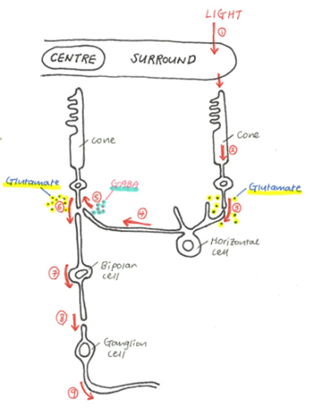
- The connection of centre cones and surround cones cones in the fovea with via acmacrine and horizontal cells.
- Antagonistic effect on the centre of receptive field.
- Occurs as the eye is trying to focus on the cencentrated (strong stimulation received in the surround) information rather than the information received in the centre which is not as concentrated as the surround information.
- Steps: On centre bipolar cells and off centre on surround ganglion is used as an example
- Presence of light in the surround receptive field and darkness in the centre receptive field.
- Hyperpolarisation of the surround cones and depolarisation of the centre cones.
- Hyperpolarisation of the surround will release less neurotransmitter (glutamate) at the synapse.
- Hence the horizontal cells are hyperpolarised.
- Stimulus on the horizontal cells will cause release of neurotransmitter called Gamma Aminobutyric Acid (GABA) at the synapse with centre cone and this will have no impact on the production of glutamate by the centre cone as hyperpolarisation of horizontal cells will not produce much of GABA.
- Large amount of glutamate is released at the synapse (due to depolarisation of the centre cones)
- On centre bipolar cells are hyperpolarised as glutamate acts as an inhibitory neurotransmitter.
- Decreased amount of glutamate is released at the synapse due to hyperpolarisation.
- This will cause hyperpolarisation of the on centre ganglion cells.
- Function of neurotransmitters:
- Glutamate
- Excitatory neurotransmitter for horizontal cell
- Inhibitory neurotransmitter for on centre bipolar cell
- Excitatory neurotransmitter for off centre bipolar cell
- Excitatory neurotransmitter for both on and off centre ganglion cell
- GABA (Gamma Aminobutyric Acid)
- Inhibitory neurotransmitter for neighbouring cones

Figure 4. The primary visual pathway. The primary visual pathway entailing the retina, optic nerves, optic chiasm, optic tract, hypothalamus, dorsal lateral geniculate nucleus, optic radiation, and primary visual cortex. Source:Dale P. (2010) Brains how they work and what they tells us about who we are, 1st edn., New Jersey: Pearson Education.
4. The signal passes through the partial crossing of axons at the optic chiasm (partial decussation) where left and right visual information delivered via the optic nerve cross to the opposite side of the lateral geniculate nucleus via the optic tract.
- The medial half (nasal half) of each retina receives light rays from the lateral portion of the visual field and crosses to the opposite lateral geniculate nucleus.
- The lateral half (temporal half) of each retina receives light rays from the medial portion of the visual field and remains to the same lateral geniculate nucleus.
- The right optic tract carries signals representing the left half of the visual field.
- The left optic tract carries signals from the right visual field.
5. All the axons synapse at the lateral geniculate nucleus and spread through the brain as the geniculostriate radiation.
6. The signal travels to the primary visual cortex in the
occipital lobe.
- Receptive fields of neuron in the primary visual cortex include two kinds of neurons:
- Simple cells
- Specific rotation axis required for visual stimulus. Non specific axis will have no stimulus.
- Receptive fields include: dark bar, light bar and light-dark bar.
- Complex cells
- Specific rotation axis of bars and edges required for visual stimulus. Non specific axis will have stimulus but less.
- Non specific to the positions of stimulatory bars and edges.
References
- Akers R.M. and Denbow D.M. (2013) Anatomy and physiology of domestic animals, 2nd edn., Iowa: John Wiley & Sons, Inc.
- Aughey E. and Frye E.L. (2001) Comparative veterinary histology, 1st edn., London: Manson Publishing Ltd.
- Dale P. (2010) Brains how they work and what they tells us about who we are, 1st edn., New Jersey: Pearson Education.
- James G.C. and Bradley G.K. (2007) Cunningham's textbook of veterinary physiology, 5th edn., St. Louis: Saunders Elsevier.
- Maggs D.J., Miller P.E. and Ofri R. (2013) Slatter's fundamentals of veterinary ophthalmology, 5th edn., Missouri: Elsevier.
- Sjaastad O.V., Sand O. and Hove K. (2010) Physiology of domestic animals, 2nd edn., Oslo: Scandinavian Veterinary Press.


