Showing posts with label cones. Show all posts

Physiology - Adaptation to light and dark

Adaptation to light and dark

Light adaptation
  • Adaptation to light > adaptation to dark
    • Recovers normal vision in less than a minute
  • Pupil constriction à restrict amount of light entering eye
  • Bleaching of photopigments
  • In bright light:
    • Sensory cells contain little uncleaved photopigment
    • Low light sensitivity
    • sensory cells are light adapted

Dark adaptation


Figure 1: Dark adaptation curves. Purple: rod adaptation curve. Green: cone adaptation curve. Red: two-stage dark adaptation curve of both rods and cones. The initial time delay between "light-adapted sensibility" and beginning of the curves is due to delay between time the lights are switched off and when measurement of curves start.
Source: Goldstein E.B. (2014) Sensation and Perception, 9th edn., Wadsworth: Cengage Learning.

  • Photopic (light-adapted) state to scotopic (dark-adapted) state
  • Maximal light sensitivity
    • Cones adapt more quickly to darkness than rods
      • Cones: 5 minutes
      • Rods: 20-30 minutes
    • Species dependent
    • Dependent on pre-existing light level
      • Brighter pre-existing light, lower rhodopsin stores à longer to reach maximal light sensitivity
  • Increase light sensitivity by
    • Dilation of pupil
    • Synaptic adaptation of retinal neurons
    • Increase rhodopsin available in rod outer segments à regeneration of rod photopigments

References
  1. Akers R.M. and Denbow D.M. (2013) Anatomy and physiology of domestic animals, 2nd edn., Iowa: John Wiley & Sons, Inc.
  2. Goldstein E.B. (2014) Sensation and Perception, 9th edn., Wadsworth: Cengage Learning.
  3. Maggs D.J., Miller P.E. and Ofri R. (2013) Slatter's fundamentals of veterinary ophthalmology, 5th edn., Missouri: Elsevier.
  4. Sjaastad O.V., Sand O. and Hove K. (2010) Physiology of domestic animals, 2nd edn., Oslo: Scandinavian Veterinary Press.

Physiology - Colour vision

Colour vision


Figure 1: Visual spectrum at different wavelengths. Humans can only perceive 390nm to 700nm on the visual spectrum. 
Source: Genoa College of Fine Arts (2014) Chromatology, Available at: http://www.accademialigustica.it/blog/?page_id=58 (Accessed: 3rd June 2014).
  • Colour of object is determined by which wavelengths of light is absorbed or reflected.
    • White objects = reflect all wavelengths
    • Black objects = absorb all wavelengths
    • Red object = absorbs more of the shortwave part of the spectrum & long wave light is reflected
  • Different type of cones with maximal sensitivity to light of different wavelengths = basis for colour vision
  • Birds & lower vertebrates: at least 3 different types of cones = EXCELLENT colour vision
  • Mammals (including all domestic animals): 2 types of cones
    • Exception: some primates & humans
  • Few species: 1 type
  • Primates (including human) : developed 3rd type
    • 3 types of cones in primates absorb light most efficiently in blue, green-yellow & orange-red parts of the spectrum
    • Blue, geen and red cones
    • Overlap between wavelengths absorbed by these 3 cones
    • Human brain can discriminate between colours
      • ie. 560nm: green & red stimulated equally & blue not affected = yellow
  • Colour vision based on 
    • 3 types of cones = Trichromatic
    • 2 types of cones = Dichromatic

Figure 2: Three different types of cones found in trichromatic primates, including humans, have different sensitivities to light of different wavelengths. Thus forming the basis for colour vision in these primates.
Source: Sjaastad O.V., Sand O. and Hove K. (2010) Physiology of domestic animals, 2nd edn., Oslo: Scandinavian Veterinary Press.

Fun facts

  • In vertebrates, it has been found that animals must discriminate between stimuli before expressing preference for a colour
    • e.g. Frogs forced to choose between two adjacent, illuminated panels. They select the bluer one
  • Fish have good colour vision
    • Fish retinas contain multiple classes of photopigments for colour vision
    • e.g Gold fish absorbency spectrum shifted to longer wavelengths
  • Primate colour vision spectral range: 400-700nm. 
  • Diurnal birds have a larger range than primates!
  • Red-green colour blindness
    • Caused by absence of long or middle wavelength-sensitive visual photopigments (specifically cones)
    • Been proven that addition of a third cone class can produce improved trichromatic vision in primates


References
  1. Genoa College of Fine Arts (2014) Chromatology, Available at: http://www.accademialigustica.it/blog/?page_id=58 (Accessed: 3rd June 2014).
  2. Jacobs G. H. (1983) 'Colour vision in animals', Endeavour, 7(3), pp. 137-140.
  3. Mancuso K., Hausworth W.W., Li Q., Connor T.B., Kuchenbecker J.A., Mauck M.C., Neitz J. and Neitz M. (2009) ' Gene therapy for red-green colour blindness in adult primates', Nature, 461, pp. 784-787.
  4. Sjaastad O.V., Sand O. and Hove K. (2010) Physiology of domestic animals, 2nd edn., Oslo: Scandinavian Veterinary Press.

Physiology - Rods and cones

Rods and cones
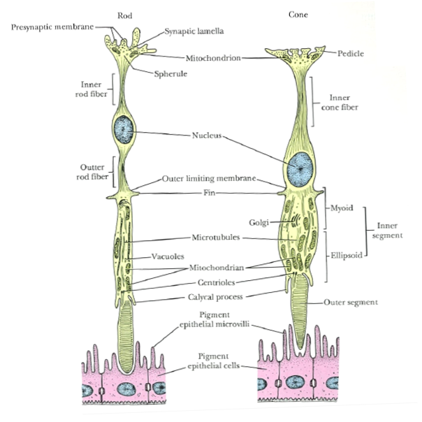
Rods and cones are the two major types of sensory cells in the eye and are located in the outer most later of the retina, closest to the choroid.


Figure 1: Diagram of rod and cone cells. Outer segments of rods and cones are closely associated with adjacent pigment epithelium.
Source: Ross M.H. and Pawlina W. (2006) Histology a text and atlas with correlated cell and molecular biology, 5th edn., Baltimore: Lippincott Williams & Wilkins.

Rods
  • Do not provide colour vision
  • Extremely sensitive
  • In poor light conditions à all vertebrae see in black, white and grey
  • In very bright conditions à lose ability to discriminate between light intensities
Cones
  • Colour vision
  • Stimulated only in good light conditions i.e. level at which rods are maximally stimulated
  • Ability to provide detailed vision in full daylight (photopic vision)
  • Area centralis of diurnal species have high number of cones
Humans

  • Density of cones are high in the fovea in the middle of the area centralis
  • Fovea in humans contain only cones à visual acuity in fovea is high
  • As fovea lack rods, area is not stimulated in weak light
  • Rod density is highest in area immediately adjacent to area centralis
Animals
  • Many species, including cattle and horses, lack a circular area centralis
  • Have visual streak
    • Density of sensory cells is high
    • Elongated region that corresponds to the projection of the horizon on retina

Table 1: Summary of the characteristics of rods and cones

Characteristics of rods and cones
Rods
Cones
Function in low light levels (scotopic)
Function in high light levels (photopic)
Sensitive to small change in light intensity
Insensitive to small change in light intensity
Low visual discrimination (low acuity)
High visual discrimination (high acuity)
Responsive to blue light
Responsive to red light
No colour differentiation
·         Contain only 1 photopigment
Colour differentiation
·         In species with 2 or more cone populations (defined by photopigments)
Sensitive to motion
Sensitive to contrast
Detect light flashing at low frequency
Detect light flashing at high frequency
More in peripheral area
More in central retina


References
  1. Maggs D.J., Miller P.E. and Ofri R. (2013) Slatter's fundamentals of veterinary ophthalmology, 5th edn., Missouri: Elsevier.
  2. Ross M.H. and Pawlina W. (2006) Histology a text and atlas with correlated cell and molecular biology, 5th edn., Baltimore: Lippincott Williams & Wilkins.
  3. Sjaastad O.V., Sand O. and Hove K. (2010) Physiology of domestic animals, 2nd edn., Oslo: Scandinavian Veterinary Press.
Anatomy - Histology of the eye

Histology of the eye

Figure 1: Tapetum fibrosum of the canine eye. 1: Sclera composed of white dense fibrous tissue 2: Lamina fusca 3: Choroid with capillaries 4: Tapetum cellulosum 5: Photosensitive layers of the retinal layers 6: Pigment layer 7: Rods and cones 8: Nuclei of rods and cones 9: Outer synaptic layer 10: Bipolar nerve cell nuclei 11: Inner synaptic layer 12: Optic nerve cells 13: Optic nerve fibres. H&E x160 Source: Aughey E. and Frye E.L. (2001) Comparative veterinary histology, 1st edn., London: Manson Publishing Ltd.


Figure 2: Tapetum fibrosum of the eye in a horse. 1: Pigment layer of the retina 2: compact layer of fibrous connective tissue 3: Choroid with blood vessels and some pigment cells. H&E. x100
Source: Aughey E. and Frye E.L. (2001) Comparative veterinary histology, 1st edn., London: Manson Publishing Ltd.

Figure 3: Posterior surface of a canine cornea. 1: Substantia propria 2: Basement membrane (Descemet's) 3: Simple squamous endothelium. H&E. x62.5
Source: Aughey E. and Frye E.L. (2001) Comparative veterinary histology, 1st edn., London: Manson Publishing Ltd.

Figure 4: Lens of a canine. 1: Anterior epithelium 2: Nuclei of lens fibres. H&E. x62.5
Source: Aughey E. and Frye E.L. (2001) Comparative veterinary histology, 1st edn., London: Manson Publishing Ltd.

Figure 5: Ciliary processes of canine. 1: Long ciliary processes extend from the ciliary body. The arrow is pointing to the epithelium 2: Ciliary muscle. H&E. x62.5
Source: Aughey E. and Frye E.L. (2001) Comparative veterinary histology, 1st edn., London: Manson Publishing Ltd.

Figure 6: Canine iris. 1: Anterior surface is covered by flattened fibrocytes 2: Core of iris is vasculature connective tissue. 3: Posterior surface epithelium is two layers of cells and part of retina. There is presence of pigments. H&E. x62.5
Source: Aughey E. and Frye E.L. (2001) Comparative veterinary histology, 1st edn., London: Manson Publishing Ltd.


Figure 7: Canine iris. Pigment in this picture is different from the pigment in figure 6. 
Source: Aughey E. and Frye E.L. (2001) Comparative veterinary histology, 1st edn., London: Manson Publishing Ltd.

Figure 8: Optic disc of a horse. 1: Scleral connective tissue 2: Optic nerve bundles. H&E. x25
Source: Aughey E. and Frye E.L. (2001) Comparative veterinary histology, 1st edn., London: Manson Publishing Ltd.


Figure 9: Layers of the retina.
Source: Maggs D.J., Miller P.E. and Ofri R. (2013) Slatter's fundamentals of veterinary ophthalmology, 5th edn., Missouri: Elsevier.

Figure 10: Fovea centralis of a monkey. FC: Fovea centralis; C: cones; Ch: Choroid; 1: Pigmented epithelium 2: Lamina of cones 3: External limiting membrane 4: Outer nuclear layer 5: Outer plexiform layer 8: Ganglion cell layers 10: Inner limiting membrane. x132
Source: Gartner L.P. and Hiatt J.L. (2009) Color atlas of histology, 5th edn., Philadelphia: Lippincott William & Wilkins.

Figure 11: Optic nerve. S: Sclera ON: Optic nerve U: Uveal tract D: Dura mater P: Pia-arachnoid A: central artery of retina.  Source: Young B. and Deaken P.J. (2002) Wheater's functional histology: A text and colour atlas, 5th edn., Edinburgh: Churchill Livingstone Elsevier .

Figure 12: Lacrimal gland separated into lobes and lobules (LO) by connective tissue (CO). SA: serous acini N: nuclei. x132 Source: Gartner L.P. and Hiatt J.L. (2009) Color atlas of histology, 5th edn., Philadelphia: Lippincott William & Wilkins.

Figure 13: Avian eye. 1: Sclera with hyaline cartilage. 2: Choroid. H. & E. x62.5.
Source: Aughey E. and Frye E.L. (2001) Comparative veterinary histology, 1st edn., London: Manson Publishing Ltd.

Figure 14: Avian eye. 1: Choroid 2: Retinal layers 3: Pigment layer 4: Rods and cones 5: Nuclei of the rods and cones 6: Outer synaptic layer 7: bipolar nerve 8: Inner synaptic layer 9: Optic nerve cells 10: Optic nerve fibres H&E. x125
Source: Aughey E. and Frye E.L. (2001) Comparative veterinary histology, 1st edn., London: Manson Publishing Ltd.

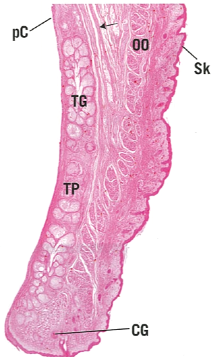
Figure 15: Eyelid. PC: Palpebral conjunctiva; OO: Orbicularis oculi; SK: Skin; TG: ; TP: ; CG: . x14
Source: Gartner L.P. and Hiatt J.L. (2009) Color atlas of histology, 5th edn., Philadelphia: Lippincott William & Wilkins.

Figure 16: Eyelid of a horse. 1: Stratified columnar epithelium with mucus secreting cells of conjunctiva 2: Sebaceous glands in the lamina propria 3: Tarsal plate. H&E. x25. Source: Aughey E. and Frye E.L. (2001) Comparative veterinary histology, 1st edn., London: Manson Publishing Ltd.


Figure 17: Nictitating membrane (third eyelid) of a canine. 1: Hyaline cartilage 2: Seromucus-secreting glands in the lamina propria. H&E. x50 Source: Aughey E. and Frye E.L. (2001) Comparative veterinary histology, 1st edn., London: Manson Publishing Ltd.

Figure 18: Nictitating membrane (third eyelid) of a horse. 1: Anterior conjunctiva surface. 2: Elastic cartilage plate. 3: Posterior conjunctival surface. 4: Lamina propria. H&E. x7.5 
Source: Aughey E. and Frye E.L. (2001) Comparative veterinary histology, 1st edn., London: Manson Publishing Ltd.

References
  1. Aughey E. and Frye E.L. (2001) Comparative veterinary histology, 1st edn., London: Manson Publishing Ltd.
  2. Gartner L.P. and Hiatt J.L. (2009) Color atlas of histology, 5th edn., Philadelphia: Lippincott William & Wilkins.
  3. Maggs D.J., Miller P.E. and Ofri R. (2013) Slatter's fundamentals of veterinary ophthalmology, 5th edn., Missouri: Elsevier.
  4. Young B. and Deaken P.J. (2002) Wheater's functional histology: A text and colour atlas, 5th edn., Edinburgh: Churchill Livingstone Elsevier .