Showing posts with label sensor. Show all posts

Physiology - Rods and cones

Rods and cones
Rods and cones are the two major types of sensory cells in the eye and are located in the outer most later of the retina, closest to the choroid.
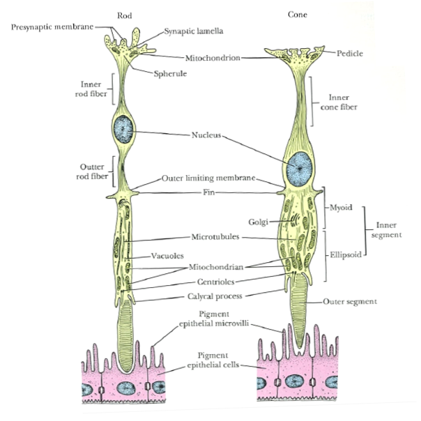

Figure 1: Diagram of rod and cone cells. Outer segments of rods and cones are closely associated with adjacent pigment epithelium.
Source: Ross M.H. and Pawlina W. (2006) Histology a text and atlas with correlated cell and molecular biology, 5th edn., Baltimore: Lippincott Williams & Wilkins.

Rods
  • Do not provide colour vision
  • Extremely sensitive
  • In poor light conditions à all vertebrae see in black, white and grey
  • In very bright conditions à lose ability to discriminate between light intensities
Cones
  • Colour vision
  • Stimulated only in good light conditions i.e. level at which rods are maximally stimulated
  • Ability to provide detailed vision in full daylight (photopic vision)
  • Area centralis of diurnal species have high number of cones
Humans

  • Density of cones are high in the fovea in the middle of the area centralis
  • Fovea in humans contain only cones à visual acuity in fovea is high
  • As fovea lack rods, area is not stimulated in weak light
  • Rod density is highest in area immediately adjacent to area centralis
Animals
  • Many species, including cattle and horses, lack a circular area centralis
  • Have visual streak
    • Density of sensory cells is high
    • Elongated region that corresponds to the projection of the horizon on retina

Table 1: Summary of the characteristics of rods and cones

Characteristics of rods and cones
Rods
Cones
Function in low light levels (scotopic)
Function in high light levels (photopic)
Sensitive to small change in light intensity
Insensitive to small change in light intensity
Low visual discrimination (low acuity)
High visual discrimination (high acuity)
Responsive to blue light
Responsive to red light
No colour differentiation
·         Contain only 1 photopigment
Colour differentiation
·         In species with 2 or more cone populations (defined by photopigments)
Sensitive to motion
Sensitive to contrast
Detect light flashing at low frequency
Detect light flashing at high frequency
More in peripheral area
More in central retina


References
  1. Maggs D.J., Miller P.E. and Ofri R. (2013) Slatter's fundamentals of veterinary ophthalmology, 5th edn., Missouri: Elsevier.
  2. Ross M.H. and Pawlina W. (2006) Histology a text and atlas with correlated cell and molecular biology, 5th edn., Baltimore: Lippincott Williams & Wilkins.
  3. Sjaastad O.V., Sand O. and Hove K. (2010) Physiology of domestic animals, 2nd edn., Oslo: Scandinavian Veterinary Press.

Physiology - Light stimulation of sensory cells

Light stimulation of sensory cells
  • When light strikes the retina, photons are trapped by the receptor molecules (rods and cones) located in the membrane discs in the outer segments
  • Membrane discs in rod = "light trap"
  • Receptor molecules in retina = photopigments
  • Rods contain photopigment, rhodopsin
  • Different types of cones have its own type of photopigments
  • Photopigments compose of opsin (protein) and retinal (produced in cell from vitamin A, retinol)
  • Photopigments are G-protein-coupled receptors


Figure 1: Cleavage of photopigments and transduction in rods and cones due to light stimulation. This causes a chemical bond change, resulting in a straightened molecule. Retinal detaches from opsin, causing conformational change in its shape. G-protein transducin binds to opsin and is activated. Hyperpolarization of the cell and reduced release of neurotransmitter ends the chain reaction. 
Source: Sjaastad O.V., Sand O. and Hove K. (2010) Physiology of domestic animals, 2nd edn., Oslo: Scandinavian Veterinary Press.

Darkness
  • Outer segment of rods and cones:
    • Concentration of cGMP in outer segment is high
    • Surface membrane:
      • High density of Na+ channels so Na+ can diffuse into cells.
      • Channels open when bound to cGMP
      • Ion channels open in dark à Na+ enters & depolarize sensory cells (light)
  • Inner segment of rods and cones:
    • Na+ pumped out via Na+-K+ pump 
    • Depolarization of inner segment keeps voltage-gated Ca2+ channels open à continuous neurotransmitter (glutamate) release from synaptic terminal of sensory cell
Light
  • When light (photons) is absorbed, 11-cis-retinal of the photopigment, rhodopsin, changes its conformational shape to all-trans-retinal
  • All-trans-retinal leaves photopigment, causing a conformational change of the photopigment
  • Now, there is a exposed binding receptor site on photopigment
  • G-protein transducin can now attach to the photopigment and is activated
  • This protein structure activates the enzyme, phosphodiesterase
  • Phosphodiesterase hydrolyses cGMP resulting in
  • closure of Na+ channels 
  • Na+ concentration in cell decreases and membrane potential = -ve
  • Rods & cones hyperpolarize
  • Release of neurotransmitter, Glutamate, is reduced
  • Results in inhibition & stimulation of ganglion cells
Figure 2: Ion currents through rods and cones in darkness. Na+ diffuses into the outer segment through open ion channels while Na+ is pumped out via Na+K+ pump in the inner segment. Light closes the ion channels.
Source: Sjaastad O.V., Sand O. and Hove K. (2010) Physiology of domestic animals, 2nd edn., Oslo: Scandinavian Veterinary Press.

Notes
  • Single photon can close many hundreds of ion channels
  • Prevents more than a million Na+ from entering cell
  • Single photon evokes detectable receptor potential in cell
Reference
  1. Sjaastad O.V., Sand O. and Hove K. (2010) Physiology of domestic animals, 2nd edn., Oslo: Scandinavian Veterinary Press.